Tethered Lipid Bilayer Membranes Assembly on Gold Electrode Surface
Tethered Lipid Bilayer Membranes Assembly on Gold Electrode Surface
Xi Wang
Tethered lipid bilayer membranes (tLBM) at the interface between a substrate and an aqueous phase are important as a novel biological membrane mimics system but also offer potential for practical applications such as biosensors. Compared to supported lipid bilayer membranes (LBMs) of which LBM sets directly on solid support, tLBM system not only guarantees a mechanically and chemically robust attachment of the lipid layer to the support but also lifts LBM away from the support so as to allow the bilayer to remain in a fluid state as a better cell membrane mimics. We used template-stripping method to fabricate pristine and atomically flat gold substrate suitable for low defect density molecular assembly, which is also compatible for large scale fabrication of electrically addressable atomically flat metal arrays.
1. Chemical Assembly of Tethered Lipid Bilayer Membrane on Gold:
Although vesicle fusion to form model membranes on hydrophilic surfaces such as glass or quartz has been widely reported, vesicles do not spontaneously rupture on Au to form LBMs without self-assembled monolayers (SAMs) on the surface. Thus, we designed lipid vesicles with functional chemical groups to promote vesicle fusion and tLBM formation on template-stripped gold (TS Au) surfaces. Vesicles are composed of lipid mixtures of 2.5 mol% of 1,2-distearoyl-sn-glycero-3-phosphoethanolamine-poly(ethylene glycol)-2000-N-[3-(2-pyridyldithio) propionate] (DSPE-PEG-PDP) / 97.5 mol% 1-palmitoyl-2-oleoyl-sn-glycero-3-phosphocholine (POPC). The disulfide group in DSPE-PEG-PDP could chemically link to Au via forming Au-thiolate bonds, which induces strong interaction between vesicles and TS Au to promote vesicle fusion on Au. The ratio of tethered lipids (DSPE-PEG-PDP) to untethered lipids (POPC) can be varied to obtain stable membranes with sufficient fluidity to mimic cellular membranes.
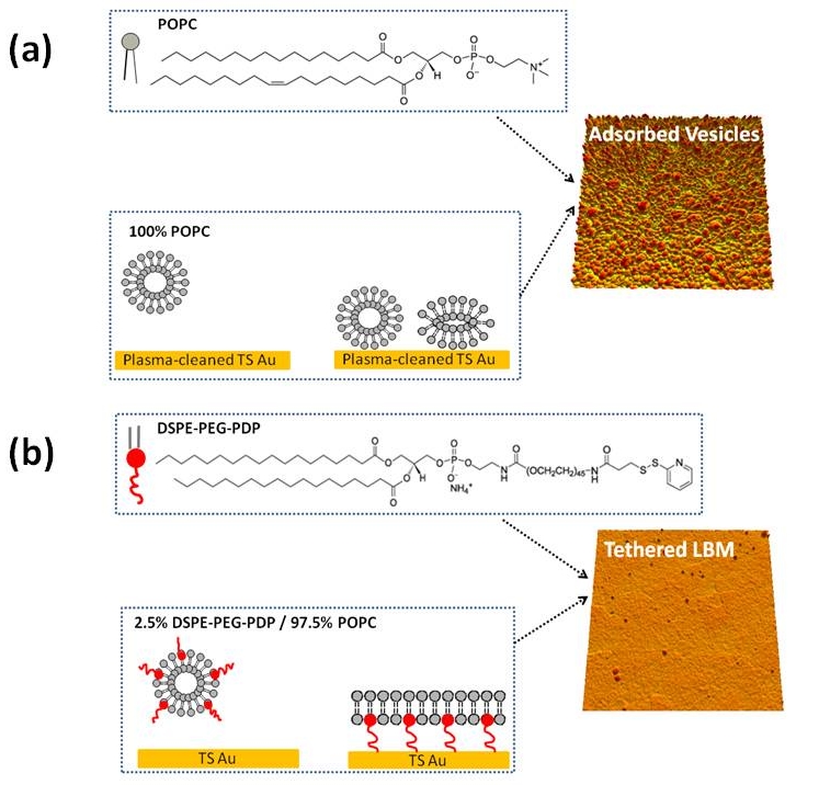
Figure 1. Schematic illustration of (a) 100% POPC vesicle interaction with plasma cleaned TS Au. Vesicles adsorbed and kept intact on plasma cleaned TS Au. (b) 2.5% DSPE-PEG-PDP / 97.5% POPC vesicle interaction with TS Au. Vesicles ruptured and formed tLBMs on TS Au.
2. Atomic Force Microscopy Characterization of tLBMs:
Atomic force microscopy (AFM) has become pivotal in visualizing biological materials as a nondestructive technique that provides nanometer scale resolution under aqueous environment and physiological conditions. Besides, AFM force spectroscopy provides pico-Newton force sensitivity to study probe-sample interactions. Force-distance curves allow one to measure local physical properties and interaction forces.
In this study, tapping mode AFM was utilized to obtain topography images of tLBMs in buffer. Force-distance curves were also acquired in fluid under contact mode to confirm the presence of tLBM on TS Au. Interpretation of the force-distance curves provides information about AFM probe-sample interaction. Specifically, there is a breakthrough event (Figure 2. Point C-D) associated with the approaching curve, and the breakthrough distance is relevant to the membrane thickness. In addition, the yield threshold force to break through the membrane indicates the membrane stability.
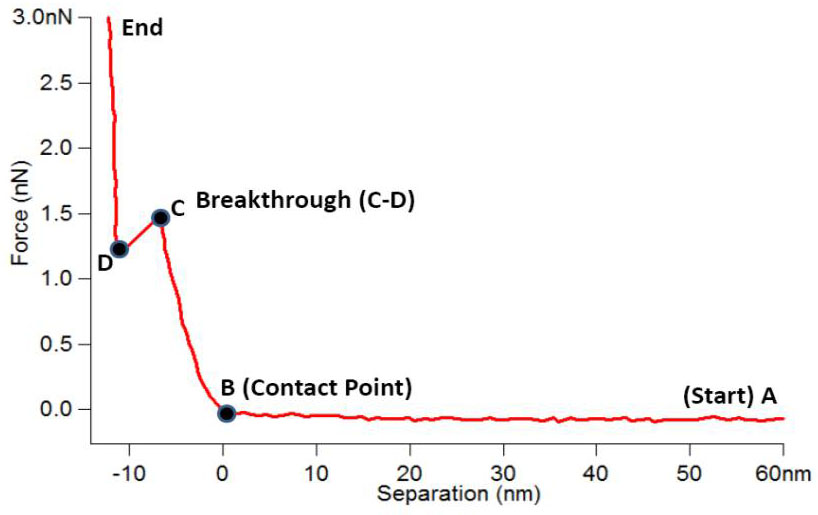
Figure 2. Typical force-distance curve of tLBM on TS Au.
Combination of AFM topography images (Figure 3. a-c) and force-distance curves (Figure 3. d-e) demonstrated that tLBMs formed on TS Au after TS Au exposure to 2.5% DSPE-PEG-PDP / 97.5% POPC, while no membrane formed after TS Au exposure to 2.5% DSPE-PEG / 97.5% POPC. Thus, the ‘PDP’ functional group for Au-thiolate bonding formation is critical for tLBMs formation on Au.
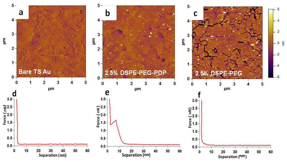
Figure 3. AFM topography images of (a) bare TS Au, (b) TS Au after incubation with vesicles composed of 2.5% DSPE-PEG-PDP / 97.5% POPC, and (c) TS Au after incubation with vesicles composed of 2.5% DSPE-PEG / 97.5% POPC. Panels (d), (e) and (f) are typical force-distance curves obtained from the scanning regions shown in (a), (b) and (c), respectively.
Our work demonstrated that tLBMs can be assembled on unfunctionalized TS Au surfaces by incorporating a disulfide functionalized lipid group in low quantities in vesicles prior to vesicle fusion. Our successful fabrication of tLBMs on TS Au via a simple one-step approach process eliminates the need for deposition of a self-assembled monolayer on Au before lipid bilayer formation, provides a robust membrane system compatible of electrical measurement of membrane-protein interactions, and has great potential to form membrane arrays by lithography patterning of TS Au.